Home
/ Plant Cell Diagram With Pencil - ComicLife.com | A blog about Comic Life in your life ... / Plant cells have a rigid, protective cell wall made up of polysaccharides.
Plant Cell Diagram With Pencil - ComicLife.com | A blog about Comic Life in your life ... / Plant cells have a rigid, protective cell wall made up of polysaccharides.
Plant Cell Diagram With Pencil - ComicLife.com | A blog about Comic Life in your life ... / Plant cells have a rigid, protective cell wall made up of polysaccharides.. The animal cell structure is the most prominent in human cheek cells. The science of biology , 4th edition, by sinauer associates ( www.sinauer.com ) and wh freeman ( www.whfreeman.com ), used with permission. On the student data sheet, color the diagram to illustrate the color bands on the chromatogram. In higher plant cells, that polysaccharide is usually cellulose. Connect the pencil leads of to dry batteries, and pass an electric current for about 20 seconds.
("u" is represents the unknown amino acid mixture). Plant cells have a rigid, protective cell wall made up of polysaccharides. Image from purves et al., life: Measure the distance from the first pencil line to the solvent front. It is important that for each tissue type you understand:
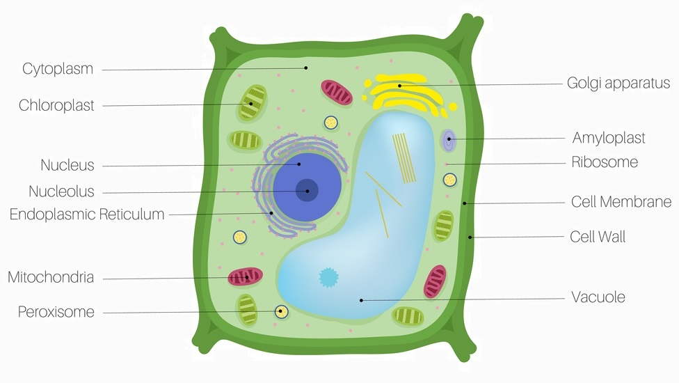
Draw the neat diagram of.plant cell. Label the following ... from hi-static.z-dn.net Cell wall (plant cells only): If examining a plant cell , tap water can be used. Diagram of a typical plant, showing the inputs and outputs of the photosynthetic process. The cell wall provides and maintains the shape of these cells and serves as a protective barrier. If examining an animal cell , physiological saline (or contact lens solution) must be used, because if plain water is used, the cell will explode from osmotic pressure. The onion skin cells were positioned beside each other (length touching length, width touching width) and formed a checkered pattern. The animal cell structure is the most prominent in human cheek cells. In this case, you can imagine that the solution is less concentrated than the cell's cytoplasm, causing water from the solution to flow into the cell.
In higher plant cells, that polysaccharide is usually cellulose.
If a solution is hypotonic to a cell, then the cell will swell when placed in the hypotonic solution. If examining an animal cell , physiological saline (or contact lens solution) must be used, because if plain water is used, the cell will explode from osmotic pressure. If a cell shrinks when placed in a solution, then the solution is hypertonic to the cell. ("u" is represents the unknown amino acid mixture). The surface of a pencil lead is rubbed lightly by the sandpaper. Label the band that traveled the greatest distance 1, the next 2, the next 3. Two leads of the pencil are passed through two holes. Describe the color of each band in data table 1, column b. The diagram above depicts how several cells adapted for the same function work in conjunction to form tissues. On the student data sheet, color the diagram to illustrate the color bands on the chromatogram. Connect the pencil leads of to dry batteries, and pass an electric current for about 20 seconds. In higher plant cells, that polysaccharide is usually cellulose. And two types of cell division:
This type of cell division is used when an organism grows. Two leads of the pencil are passed through two holes. If examining an animal cell , physiological saline (or contact lens solution) must be used, because if plain water is used, the cell will explode from osmotic pressure. In higher plant cells, that polysaccharide is usually cellulose. Describe the color of each band in data table 1, column b.
Plant Cell - Definition, Parts and Functions | Biology ... from biologydictionary.net Continue until all bands are labeled. Along this line ten light crosses ("x") are marked at intervals of about 2 cm. Measure the distance from the first pencil line to the solvent front. The diagram above depicts how several cells adapted for the same function work in conjunction to form tissues. If examining a plant cell , tap water can be used. Diagram of a typical plant, showing the inputs and outputs of the photosynthetic process. The onion skin cell, an example of a plant cell, generally has a rigid, rectangular shape. The animal cell structure is the most prominent in human cheek cells.
It is important that for each tissue type you understand:
The surface of a pencil lead is rubbed lightly by the sandpaper. Figure 4.2 provides an overview of the types of plant tissues being studied in this chapter. If a solution is hypotonic to a cell, then the cell will swell when placed in the hypotonic solution. If examining an animal cell , physiological saline (or contact lens solution) must be used, because if plain water is used, the cell will explode from osmotic pressure. Diagram of a typical plant, showing the inputs and outputs of the photosynthetic process. The onion skin cells were positioned beside each other (length touching length, width touching width) and formed a checkered pattern. Fluid collects in the plant cell vacuole and pushes out against the cell wall. Label the band that traveled the greatest distance 1, the next 2, the next 3. Describe the color of each band in data table 1, column b. Measure the distance from the first pencil line to the solvent front. The animal cell structure is the most prominent in human cheek cells. In higher plant cells, that polysaccharide is usually cellulose. Cell wall (plant cells only):
Cell wall (plant cells only): The surface of a pencil lead is rubbed lightly by the sandpaper. In higher plant cells, that polysaccharide is usually cellulose. This type of cell division is used when an organism grows. And two types of cell division:
Graphique d'éducation de biologie pour le diagramme de ... from i.pinimg.com On a clean sheet of chromatography paper with size about 12 cm by 22 cm, a light pencil line is marked to the bottom and about 1.5 cm away. Measure the distance from the first pencil line to the solvent front. If a cell shrinks when placed in a solution, then the solution is hypertonic to the cell. The liquid used depends on the type of cell being viewed: Two leads of the pencil are passed through two holes. If examining a plant cell , tap water can be used. Along this line ten light crosses ("x") are marked at intervals of about 2 cm. The surface of a pencil lead is rubbed lightly by the sandpaper.
This type of cell division is used when an organism grows.
On a clean sheet of chromatography paper with size about 12 cm by 22 cm, a light pencil line is marked to the bottom and about 1.5 cm away. Measure the distance from the first pencil line to the solvent front. ("u" is represents the unknown amino acid mixture). And two types of cell division: The diagram above depicts how several cells adapted for the same function work in conjunction to form tissues. If examining a plant cell , tap water can be used. On the student data sheet, color the diagram to illustrate the color bands on the chromatogram. The science of biology , 4th edition, by sinauer associates ( www.sinauer.com ) and wh freeman ( www.whfreeman.com ), used with permission. The animal cell structure is the most prominent in human cheek cells. If a solution is hypotonic to a cell, then the cell will swell when placed in the hypotonic solution. Cell wall (plant cells only): It is important that for each tissue type you understand: Connect the pencil leads of to dry batteries, and pass an electric current for about 20 seconds.
Share :
Post a Comment
for "Plant Cell Diagram With Pencil - ComicLife.com | A blog about Comic Life in your life ... / Plant cells have a rigid, protective cell wall made up of polysaccharides."


Post a Comment for "Plant Cell Diagram With Pencil - ComicLife.com | A blog about Comic Life in your life ... / Plant cells have a rigid, protective cell wall made up of polysaccharides."